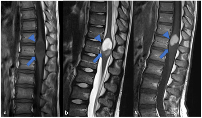
Figure 2.
Pilocytic astrocytoma in an eight-year-old child, including expansive mass and distinct solid cystic components at level D11-12. Sagittal T1-weighted (a), T2-weighted (b), and post-contrast T1-weighted (c) images demonstrate a cystic-like cranial component with evident and homogeneous enhancement (arrowheads) and a caudal solid component without enhancement (arrows).

