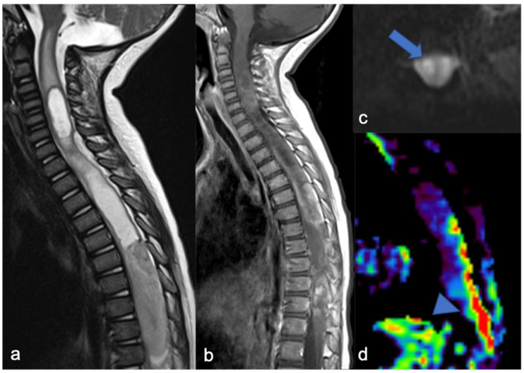
Figure 4.
Sagittal T2-weighted image (a), post-contrast T1-weighted (b), DWI (c; T10-T11) and DSC (d). High-grade glioma with cervical–thoracic epicenter and holocordal involvement of the spinal cord in a two-year-old child. The neoplasm is characterized by inhomogeneous enhancement. Components with restricted diffusion (arrow) and increased rCBV (arrowhead) are shown.

