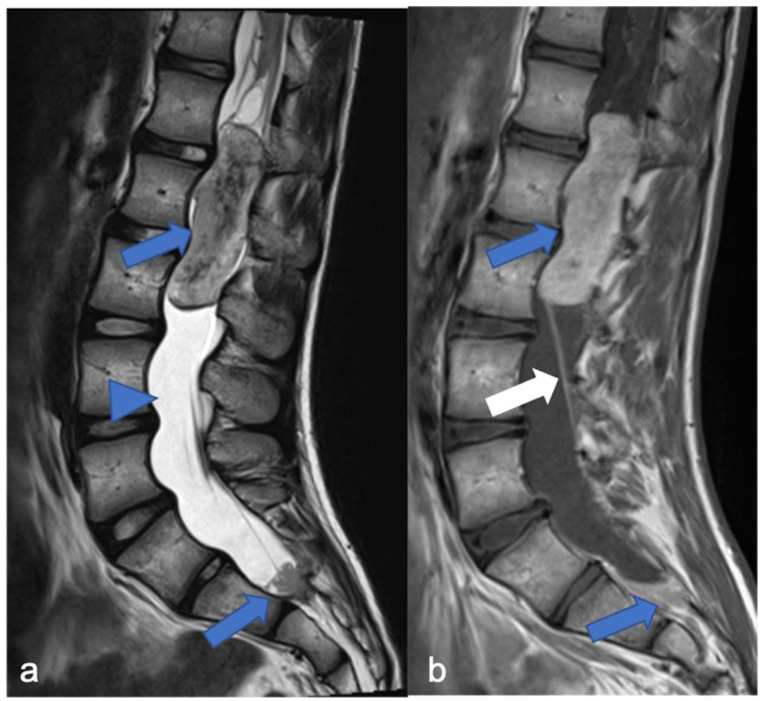
Figure 8.
Myxopapillary ependymoma in a fourteen-year-old child located in the lumbo-sacral region. Sagittal T2-weighted (a) and post-contrast T1-weighted (b) images demonstrate solid cranial and caudal enhancing components (blue arrows) and a pseudocystic non-enhancing component (arrowhead). The detail of the filum terminale is also highlighted (white arrow).

