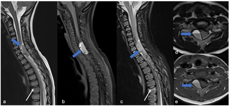
Figure 15.
Tumor-like appearance of a spinal “formation” in a six-year-old child. MRI was performed after a traumatic event. Sagittal T2-weighted (a), fat suppression T1-weighted (b), fat suppression T2-weighted (c), axial T2-weighted (d), and T1-weighted (e) images demonstrate intradural-extramedullary cystic-like formation with a fluid–fluid level (blue arrows) and spinal cord dislocation. Traumatic deformation of the superior vertebral plateau is also evident (white arrows). The definitive diagnosis was post-traumatic pseudomeningocele.

