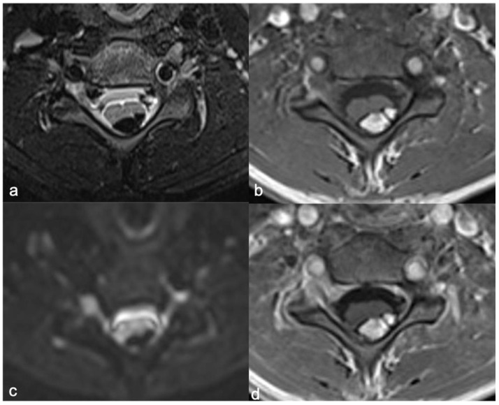
Figure 16.
Intradural-extramedullary cervical expansive lesion in a ten-year-old child. Axial fat suppression T2-weighted (a), T1-weighted (b), DWI (c), fat suppression post-contrast T1-weighted images (d) demonstrate hyperintense lesion (T1 images), hypointense in fat suppression T2-w image, without diffusion restriction and enhancement. The diagnosis of lipoma is clear.

