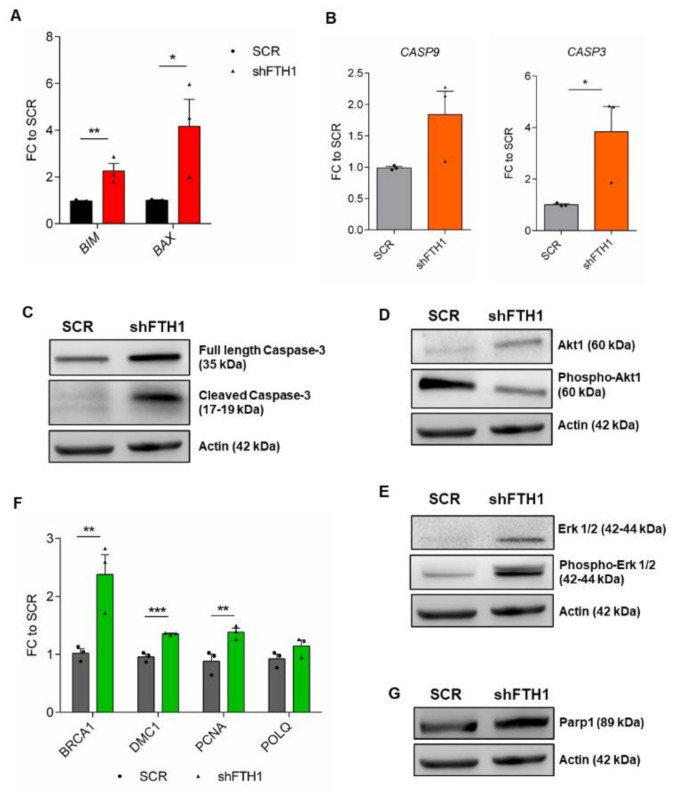
Figure 4.
FTH1 silencing leads to expression of apoptosis and DNA damage markers in hESCs. (A,B) qRT-PCR for apoptosis-associated genes BIM and BAX (A) and CASP3 and CASP9 (B) showing increased expression levels of apoptotic marker in FTH1-KD hESCs compared to SCR control; (C) Activation of apoptotic signaling in FTH1-KD hESCs is demonstrated also by immunoblot analysis for full length and cleaved form of caspase-3; (D) Western blot analysis of Akt1 and phosphorylated Akt1 (Ser473, pAkt) protein expression; (E) Immunoblot for total Erk1/2 and phosphorylated Erk1/2 (Thr202/Tyr204, pErk1/2) in SCR control and FTH1-KD hESCs; (F) qRT-PCR analysis for DNA damage related genes (BRCA1, DMC1, PCNA, POLQ) in control and silenced hESCs; (G) Western blot analysis for Parp1 protein expression in SCR and FTH1-silenced cells. For all Western blots shown, actin levels were evaluated as loading control. Results of qRT-PCR were normalized to measurements in SCR cells (n = 3); * p ≤ 0.05; ** p ≤ 0.01, t-test, *** p ≤ 0.001. Small dots refer to SCR replicates, while triangles refer to shFTH1 triplicates.

