Figure 5.
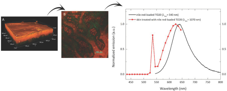
Porcine skin treated with nile red-loaded TPGS micelles. (A) Volume rendering of flat porcine skin reconstructed from the Z-stack (Z-step: 0.8 μm, total depth: 151 μm); (B) intermediate region between epidermis and dermis, acquired about 80 μm from the sample surface; dermis papillae are evident (in green the collagen) together with annexial, vascular and corpuscular elements; (C) comparison between the normalized emission spectrum of an aqueous suspension of nile red-loaded TO micelles (black line) and the emission spectrum acquired in the skin region shown in (B) (red line). The sharp peaks at 535 nm (red line) is relevant to the SHG signal. All the images have been acquired with excitation wavelength of 1070 nm. A more complete and detailed view of the flat porcine skin 3D reconstruction is available in Supplementary Movie S1.

