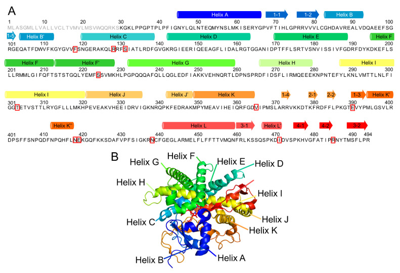
Figure 1.
Numbering of the secondary structures of CYP2A6. (A) Correspondence between the amino acid sequence and secondary structures. The circular column and arrow indicate helix and β-strand formation regions, respectively. The gray letters of the amino acid sequence are the disordered region in the crystal structure. The mutation sites relevant to 10 variants investigated in this study are shown in red squares. (B) Correspondence between the three-dimensional structure and secondary structures.

