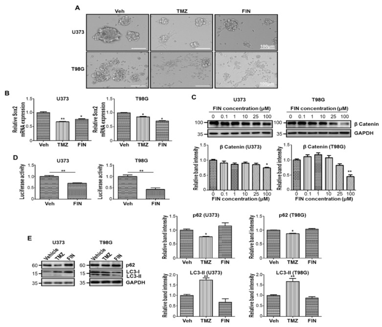
Figure 1.
Glioblastoma stem-like cells are sensitive to high-dose finasteride. (A) Cell morphology of U373 (upper) and T98G (lower) glioblastoma cells under sphere-forming culture condition upon treatment of DMSO or temozolomide (TMZ) or 100 μM finasteride (FIN) for 24 h. (B) The mRNA expression of Sox2 in U373 (left) and T98G (right) cells upon vehicle or drug treatment, as measured by quantitative RT-PCR analysis. The mRNA level of vehicle-treated cells was set to 1. (C) Immunoblot analysis of β-catenin in U373 (left) or T98G (right) cells after treatment with vehicle or FIN at the indicated concentration for 24 h. GAPDH was used as a loading control. The relative band intensities of β-catenin are shown below. The intensities of vehicle-treated samples were arbitrarily set to 1. (D) β-catenin activity was measured in U373 (left) or T98G (right) cells by TOPFlash reporter assay upon treatment of DMSO or 100 μM FIN for 24 h. (E) Immunoblot analysis of P62 and LC3 in U373 (left) or T98G (right) cells after vehicle or drug treatment for 24 h. The relative band intensities of P62 and LC3-II are represented. The intensities of vehicle-treated samples were arbitrarily set to 1. * p < 0.05, ** p < 0.01.

