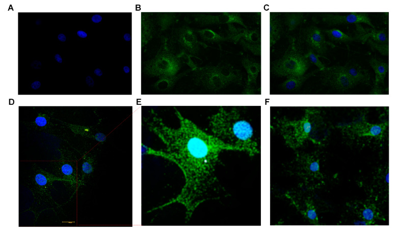
Figure 4.
Immunocytochemistry studies demonstrating expression of taste-specific protein markers in cultured BTBCs. (A–C) Confocal images of cultured BTBCs expressing α-gustducin; (A) DAPI stained nucleus (blue), (B) Alexa Fluor 488 staining of the protein α-gustducin and (C) combined pictures. (D) PLCβ2 expressing cells and (E) the inset displays magnified view of the region on the red box; and (F) cells expressing bitter receptor T2R7.

