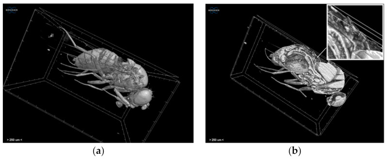
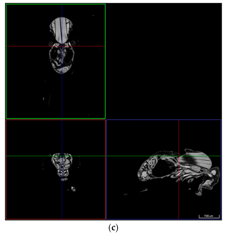
Figure 12.
Volume rendering of (a) the whole specimen and (b) the virtually dissected specimen and (c) transaxial, sagittal, and coronal images of the Drosophila melanogaster acquired through the Skyscan 1172 micro-CT scanner. The white arrow and the cross-point of the coloured lines indicate the heart position.


