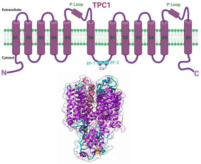
Figure 6.
Putative structure of TPC1 channel. (Top) Schematic cartoon representation of individual plant TPC1 channel subunit comprising two repeated domains showing six membrane-spanning regions (S1–S6), two pore loops (P), and joined via a cytosolic linker containing two Ca2+ binding EF-hands (EF1 and EF2). (Bottom) TPC1 secondary 3D structure model showing two subunits in transparent surface view was developed from PDB structure 5DQQ (Arabidopsis thaliana TPC1). The image was prepared using Chimera software [122]. Created with BioRender.com (accessed on 30 August 2021).

