Figure 4.
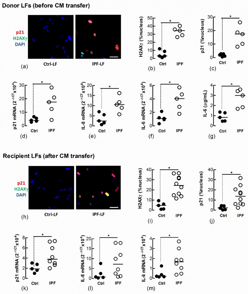
Conditioned medium from IPF-LFs induces senescence in naïve Ctrl-LFs. Sub-confluent naïve recipient Ctrl-LFs were exposed to CM from Ctrl- or IPF-LFs for 3 d (n = 5–10). (a–g) Levels of senescence markers in donor Ctrl- and IPF-LFs prior to culture with CM. Representative immunofluorescence images from Ctrl- and IPF-LF cultures show nuclear localisation of H2AXγ and p21 (blue = DAPI, red = p21 and green = H2AXγ). (h–m) Levels of senescence markers in naïve Ctrl-LFs 3 d after culture with CM. Each data point for the IPF treatment group represents a unique combination of CM from a “donor” IPF patient and recipient Ctrl-LF. CM from LFs of five separate IPF patients were used. (* p < 0.05). Scale bar = 50 μm.

