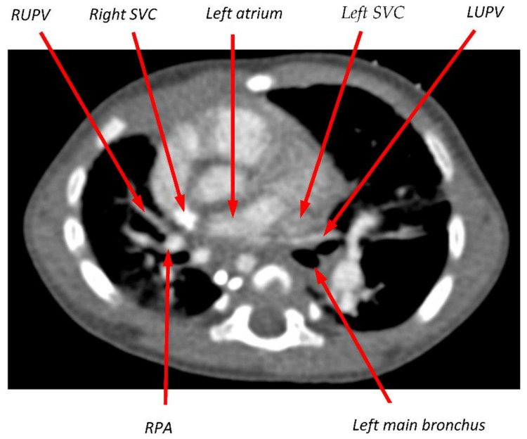
Figure 3.
Transverse slice of CT scan from Pt2 at the level of the left atrium (LA) and upper pulmonary veins. The RUPV was documented as atretic at the time of this scan, but its course had presumably been between the right superior vena cava (SVC) and the right pulmonary artery (RPA). On the left side, the takeoff of the LUPV from the LA is sharply angulated, and the vein appears compressed as it passes between the left SVC and the left main bronchus.

