Figure 4.
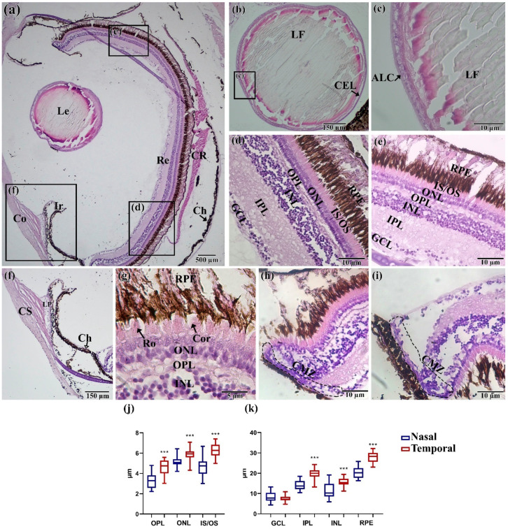
Histological structure of the eye of N. guentheri: (a) Total cut of the eye; (b,c) lens; (d) temporal part of the retina; (e) nasal part of the retina; (f) cornea and iris; (g) layer of receptor cell; (h,i) ciliary marginal zone; (j,k) thickness of layers in the temporal and nasal areas of the retina (n = 3 × 50). The value (***—p < 0.001) from one-way ANOVA with comparison using Tukey’s post hoc analysis. Le—lens, Re—retina, Co—cornea, Ir—iris, CR—choroid rete, CS—corneal stroma, Ch—choroid, GCL—ganglion cell layer, IPL—inner plexiform layer, INL—inner nuclear layer, OPL—outer plexiform layer, ONL—outer nuclear layer, IS/OS—inner segment/outer segment of the photoreceptor cells, RPE—retinal pigment epithelium/cells, LF—lens fibers, ALC—acellular lens capsule, CEL—cuboidal epithelial lens cells; LP—ligamentum pectinatum; Ro—rods; Con—cones; CMZ—ciliary marginal zone.

