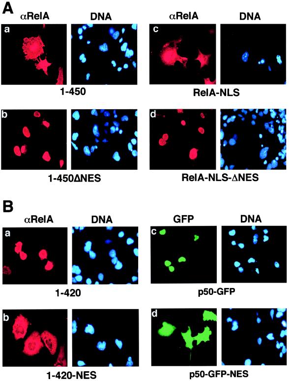
FIG. 3.
The NES-like sequence of RelA promotes cytoplasmic localization of RelA as well as p50. (A) COS cells were transfected with cDNA expression vectors encoding the RelA proteins indicated below each of the panels. The cells were stained with anti-RelA plus Texas red-conjugated rabbit IgG (αRelA), counterstained with Hoechst, and visualized as described in the legend for Fig. 1B. (B) COS cells were transfected with cDNA expression vectors encoding the proteins indicated below the panels. The cells were stained with an anti-RelA antiserum and Texas red-conjugated rabbit IgG. The expression of the RelA mutants (a and b) was visualized with a rhodamine filter (αRelA), whereas the expression of p50-GFP fusion proteins (c and d) was visualized via the autofluorescence of GFP by using an FITC filter (GFP). The nuclei of the cells were visualized by DNA staining with Hoechst (DNA).

