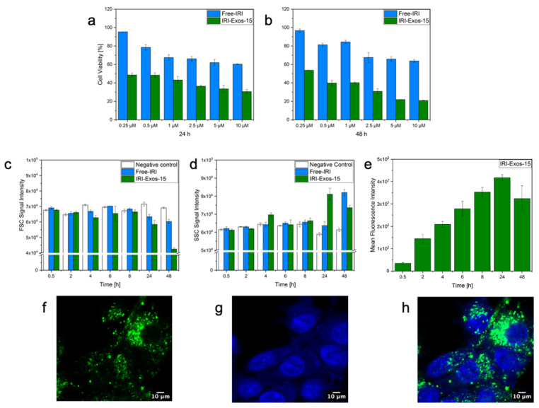
Figure 4.
MTT Cytotoxicity assay of IRI-Exos-15 and Free-IRI cultured on U87-MG cells at 24 h (a) and 48 h (b); Flow cytometry of IRI-Exos-15 up to 48 h of incubation with U87-MG cells: (c) FSC signal, (d) SSC analysis, and (e) mean fluorescence intensity. All data are presented as mean ± s.d (n = 3). Images of Exos internalization at 24 h of incubation with U87-MG cells; (f) IRI-Exos-15 stained with PKH67; (g) U87-MG cells nuclei stained with Hoechst; and (h) merged channels.

