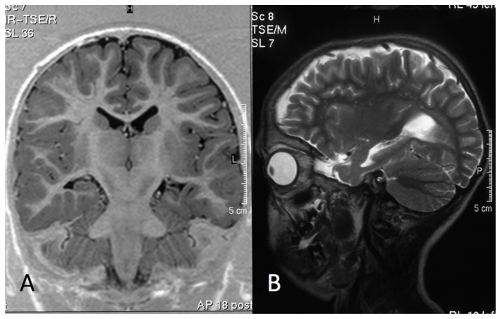
Figure 3.
Group 1: MRI of a 11-year-old boy (Pt1, Lys 601Gln). (A) Inversion Recovery (IR) Turbo Spin Echo (TSE) Image, coronal plane. (B) TSE T2 weighted image, sagittal plane. The MRI shows a small left hippocampus and enlarged temporal horn and collateral sulcus, consistent with left-sided hippocampal sclerosis. Note the mild widening of left sylvian fissure and of the left fronto-temporal cerebrospinal fluid (CSF) spaces, consistent with cortical atrophy.

