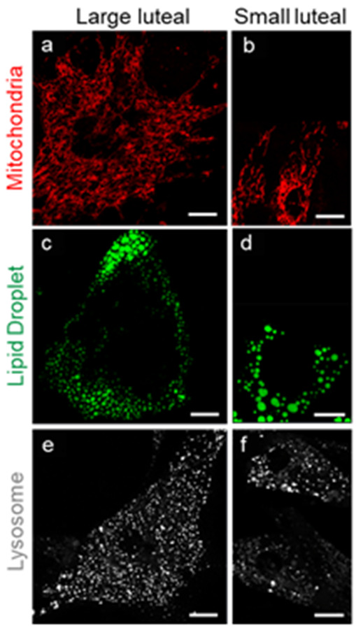
Figure 1.

Organelle differences between steroidogenic large and small bovine luteal cells. Confocal microscopy was employed to visualize intracellular organelles in cultured bovine luteal cells. Representative micrograms of mitochondria ((a,b) MitoTracker); lipid droplets ((c,d) TopFluor Cholesterol);and lysosomes ((e,f) LysoTracker) in large and small luteal cells (left to right). Micron bar represents 10 µm.
