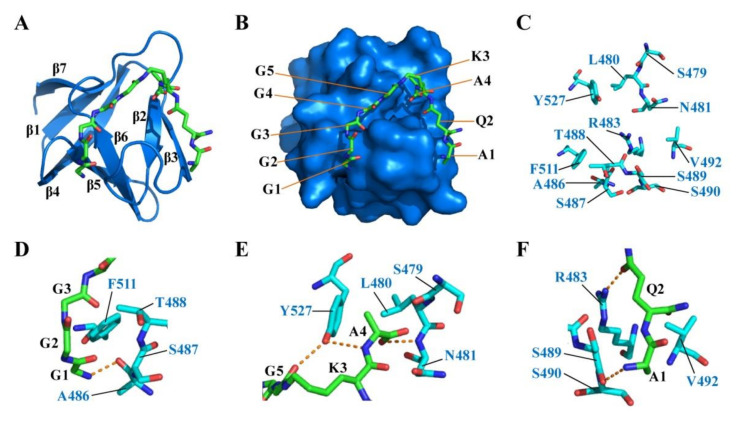
Figure 6.
Binding model of CpAcp SH3b6 with P4-G5 (K5T) peptide. (A) Complex model of CpAcp SH3b6 bound by P4-G5 with SH3b6 shown in cartoon (marine) and P4-G5 peptide in stick representation. Secondary elements of SH3b6 are marked. (B) Complex model of CpAcp SH3b6 bound by P4-G5 with SH3b6 in surface (marine) representation. Amino-acid units of P4-G5 are marked. (C) The residues of SH3b6 involved in interaction with P4-G5 are shown as sticks. (D–F) Three interacting sites between SH3b6 and P4-G5 are shown with carbon colored in cyan and green, respectively. The interacting residues are indicated. Hydrogen bonds are shown as orange dashed lines.

