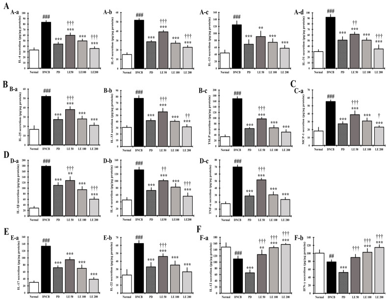
Figure 7.
Effect of LE on AD-related cytokine and chemokine secretion in NC/Nga mouse splenocytes. (A) The levels of (A-a) IL-4, (A-b) IL-5, (A-c) IL-13, and (A-d) IL-31 in the supernatant of splenocytes. (B) The levels of (B-a) IL-25, (B-b) IL-33, and (B-c) TSLP in the supernatant of splenocytes. (C) The levels of (C-a) MCP-1 in the supernatant of splenocytes. (D) The levels of (D-a) IL-1β, (D-b) IL-6, and (D-c) TNF-α in the supernatant of splenocytes. (E) The levels of (E-a) IL-17 and (E-b) IL-22 in the supernatant of splenocytes. (F) The levels of Th1-mediated cytokines (F-a) IL-12 and (F-b) IFN-γ. Splenocytes from NC/Nga mice were stimulated with Con-A for 72 h, and then the supernatant was measured using ELISA. Cytokines and chemokines were normalized to the protein concentration of the lysate. The results were expressed as means ± SD (n = 6). ## p < 0.01, ### p < 0.001 vs. normal (DNCB untreated group), ** p < 0.01, *** p < 0.001 vs. DNCB (negative control; DNCB treated group), PD (positive control; prednisolone 3 mg/kg) treatment group, LE (LE 50, 100 or 200 mg/kg) treatment group, and † p < 0.05, †† p < 0.01, and ††† p < 0.001 vs. PD (positive control; prednisolone 3 mg/kg) treatment group.

