Figure 5.
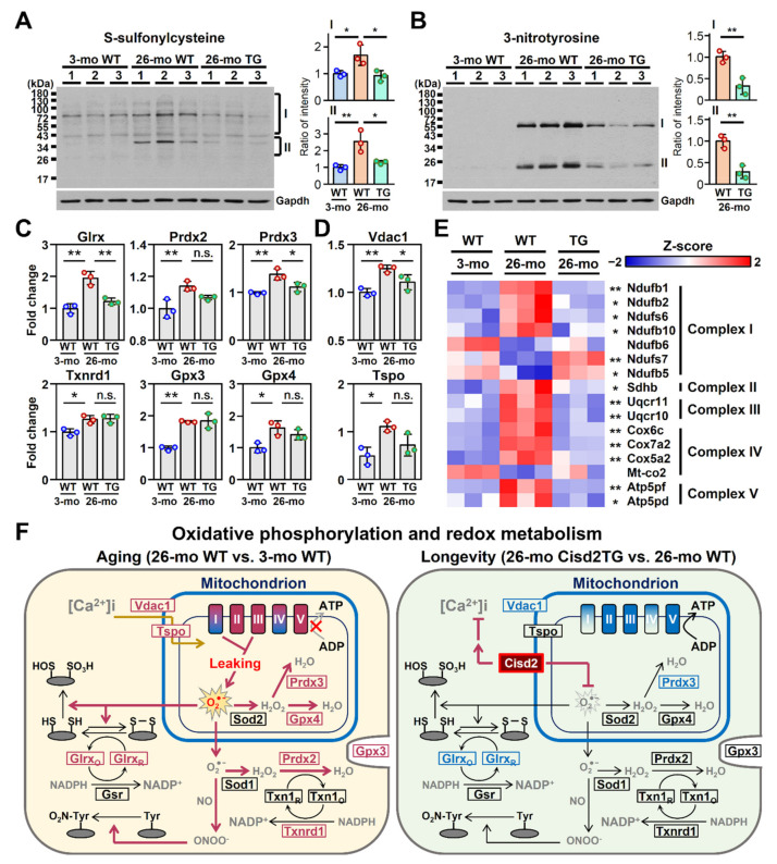
Cisd2 modulates redox status, calcium homeostasis and mitochondrial respiration. (A,B) Western blot analyses of the oxidative modification of liver proteins by S-sulfonylcysteine and 3-nitrotyrosine in the 3-mo WT, 26-mo WT and 26-mo Cisd2TG mice. Quantification of each sample is based on the entire intensities of each lane or region normalized against GAPDH; * p < 0.05; ** p < 0.01 (Tukey’s HSD test after the quantification of S-sulfonylcysteine and Student’s t-test after the quantification of 3-nitrotyrosine). (C,D) The aging-associated DEPs involved in calcium transport and the antioxidant response. The data are represented as the means ± SD; * p < 0.05; ** p < 0.01 (Tukey’s HSD test after the one-way ANOVA had reached statistical significance). (E) A heatmap revealing the difference between the 26-mo Cisd2TG and 26-mo WT mice in the aging-associated DEPs related to complexes I, II, III, IV and V of the mitochondrial ETC; * p < 0.05; ** p < 0.01 (Tukey’s HSD test). (F) A schematic illustration of redox status, calcium homeostasis and mitochondrial oxidative phosphorylation of the naturally aged WT mice and the long-lived Cisd2TG mice. Red indicates upregulation and blue indicates downregulation of the relevant proteins. n.s., non-significant. The “x” denotes the inhibited ATP synthesis during aging compared to the 26-mo Cisd2TG mice. The gray arrow was also used to emphasize this decreasing trend.

