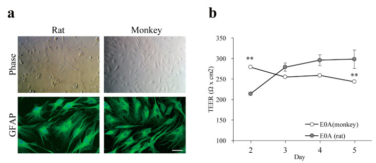
Figure 3.
(a) Characterization of primary monkey astrocytes by immunofluorescence microscopy. Both monkey and rat astrocytes express glial fibrillary acidic protein (GFAP). Cell nuclei were counterstained with TO-PRO-3 (blue). Bar = 50 μm; (b) Barrier function of the co-culture BBB models with astrocytes. BECs from monkey were co-cultured with astrocytes from either monkey (E0A (monkey)) or rat (E0A (rat)). Barrier function was assessed by measuring TEER. Values presented are means ± SEM. (n = 3, ** p < 0.01). E00: monkey brain endothelial monolayers; E0A: co-culture of monkey brain endothelial cells and astrocytes (rat or monkey).

