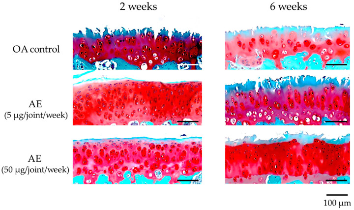
Figure 4.
Histologic appearances of medial femoral condyle cartilage (safranin O staining) of the right knee joint of experimental OA model rats. Samples were obtained at 2 and 6 weeks after surgery from the joints without treatment (OA control, n = 18), and with lower dose AE treatment (5 µg/joint/week, n = 20) and higher dose AE treatment (50 µg/joint/week, n = 19). Progression of cartilage destruction was milder in the treatment group than in the control group at 6 weeks after surgery.

