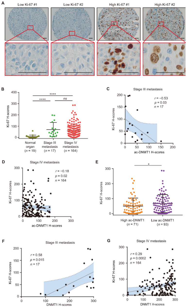
Figure 6.
Ac-DNMT1 and Ki-67 protein levels correlate in metastatic melanoma. (A,B) Representative IHC images and H-score quantification of Ki-67 in normal organ tissues, stage III, and stage IV metastatic melanomas from the TMA cohort. Scale bars = 20 μm. (C,D) Correlation between Ki-67 and ac-DNMT1 H-scores in stage III (C) or stage IV (D) metastatic FFPE tissues. (E) Comparison of Ki-67 H-scores in stage IV patients with high or low ac-DNMT1 levels. (F,G) Correlation between Ki-67 and DNMT1 IHC H-scores in stage III (F) or stage IV (G) metastatic FFPE tissues. The best-fit line (straight line) and the 95% CI (dotted line) are shown in grey. Data represent the mean ± SD. ns: not significant, * p < 0.05, and **** p < 0.0001.

