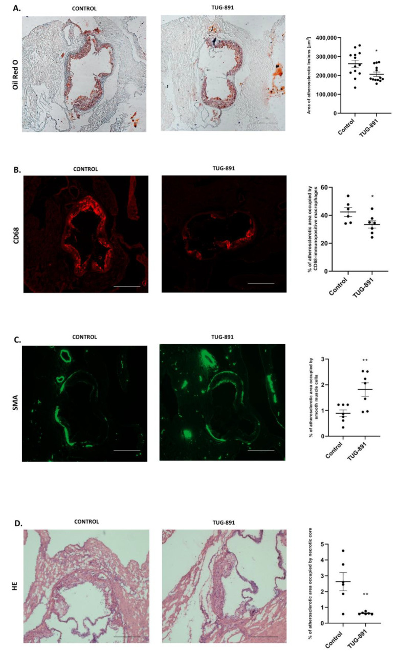
Figure 1.
(A) Representative micrographs showing Oil Red O–stained lesions in control group and TUG-891-treated group. Atherosclerotic lesions area is measured by the cross-section method in control group and TUG-891-treated group (p < 0.05; n= 13 per group). (B,C) Macrophage infiltrated in the atherosclerotic lesion of TUG-891-treated apoE−/− mice. Immunohistochemical staining of aortic roots showing CD68-positive macrophages (red), α-SMA-positive (green). (mean ± SEM; * p < 0.05 as compared to apoE−/− mice; n = 6–7 per group). (D) Content of necrotic core in the atherosclerotic lesion of TUG-891-treated apoE−/− mice. Immunohistochemical staining showing necrotic core in atherosclerotic lesions of apoE−/− mice and TUG-891-treated apoE−/− mice (mean ± SEM; ** p < 0.005; n = 6–7 per group).

