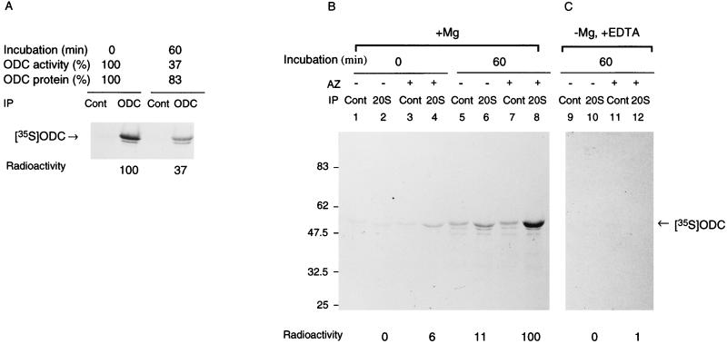
FIG. 5.
Energy- and AZ-dependent binding of ODC to the 26S proteasome. (A) ODC was incubated with a crude cell extract of HTC cells in the inactivation assay mixture for 60 min at 37°C. Aliquots of the reaction mixture before and after incubation were examined for ODC activity and ODC protein, and aliquots were immunoprecipitated with monoclonal antibody to ODC (HO101) or control IgG followed by SDS-PAGE and autoradiography. Intensities of ODC bands were determined with an image analyzer (Fujix BAS2000), and specific bindings were obtained by subtracting the value with control IgG and are expressed relative to the amount of time zero control. (B and C) ODC was incubated with partially purified 26S proteasome in the inactivation assay mixture. Where indicated, AZ was removed from the reaction mixture or EDTA was added to it instead of MgCl2. The incubated mixture was immunoprecipitated with anti-20S proteasome antibody (20S) or control IgG (Cont). The mixture without incubation was similarly treated with antibodies. The immunoprecipitates were washed extensively with buffer containing 0.1% SDS and 0.1% Triton X-100 and subjected to SDS-PAGE and then to autoradiography. Panels B and C represent separate experiments. The control (with MgCl2, without EDTA) is not shown in panel C but was almost the same as in panel B. Intensities of ODC bands were determined by an image analyzer, and the specific precipitation of ODC with anti-20S proteasome antibody was obtained by subtracting the value with control IgG and is expressed relative to the amount immunoprecipitated when ODC was incubated in the presence of AZ and MgCl2.

