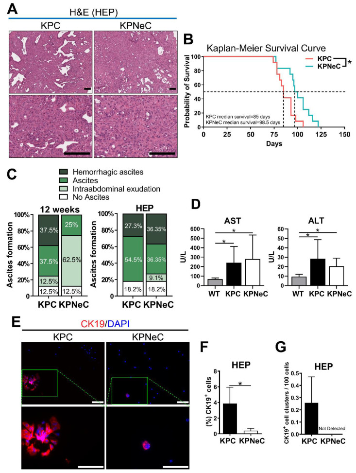
Figure 2.
Pancreas-specific NEMO ablation increases the survival rate of KPC mice. (A) H&E staining on pancreatic sections of HEP-analyzed KPC and KPNeC mice on different magnifications. Scale bar: 100 μm. (B) Kaplan–Meier survival curve for KPC (coral line) and KPNeC (cyan line) mice (n = 12 mice/group; log-rank test: * p < 0.5). (C) Grading of ascites development in 12-week-old and HEP-analyzed KPC and KPNeC mice (n = 8 mice/group). (D) Quantification of aspartate transaminase (AST) and alanine transaminase (ALT) levels in the serum of the indicated groups. (U/L = Units/Liter; serum from KPC and KPNeC mice was extracted at their HEP; serum from WT mice was extracted at the age of 12 weeks; n ≥ 7 mice/group; student’s t-test: * p < 0.5). (E) Visualization of CK19+ ascitic cells isolated from HEP-analyzed KPC and KPNeC mice. Nuclear staining with DAPI, scale bar: 100 μm. (F) Percentage of CK19+ cells to the total number of ascitic cells (n = 3 mice/group; student’s t-test: * p < 0.5). (G) Percentage of CK19+ cell clusters per 100 ascitic cells (n = 3 mice/group).

