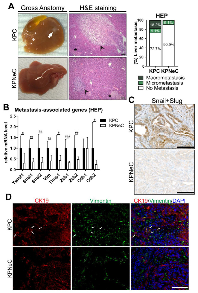
Figure 3.
Pancreas-specific NEMO ablation abrogates macro-metastasis in KPC mice and blocks epithelial–mesenchymal transition (EMT). (A) Left: Visualization of gross anatomy of the liver and H&E staining on liver sections of HEP-analyzed KPC and KPNeC mice. Arrow: Macro-metastasis. Arrowhead: Micro-metastasis. Asterisk: necrosis. Scale bar: 100 μm. Right: Quantification of liver macro- and micro-metastasis of HEP-euthanized KPC and KPNeC mice. (n = 11 mice/group). (B) Quantitative RT-PCR for the expression of the indicated transcripts in pancreatic tissue of HEP-analyzed KPC and KPNeC mice, given relative to KPC mice, which were set to 1 (n ≥ 6 mice/group; Mann–Whitney test: # p < 0.05, ## p < 0.01, ### p < 0.001). (C) Visualization of Snail+Slug staining on pancreatic sections of 12-week-old KPC and KPNeC mice. Scale bar: 100 μm. (D) Visualization of CK19+ and Vimentin+ cells on pancreatic sections of 12-week-old KPC and KPNeC mice. Nuclear staining with DAPI. Arrow: Indicative CK19+/Vimentin+ cells, scale bar: 100 μm.

