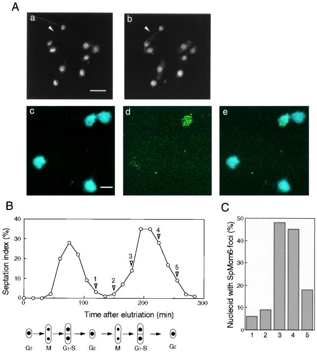
FIG. 5.
Subnuclear localization of SpMcm6p. (A) Exponentially growing strain 972 cells were fixed and stained with DAPI (a) or immunostained with affinity-purified anti-SpMcm6 antibody and FITC-conjugated anti-rabbit IgG (b). A binuclear cell in post-M to S phase is indicated with an arrowhead in parts a and b. Spread nucleoids prepared as described in Materials and Methods were stained with DAPI (c) or immunostained with anti-SpMcm6 antibody (d). Merged images of DAPI and immunostaining are shown in part e. Bars, 5 μm. (B) Small G2 phase cells collected by centrifugal elutriation were incubated in YPD at 30°C. The population of cells with septums (septation index) was measured every 15 min. (C) At the indicated time points (points 1 to 5, shown by arrowheads), spread nucleoids were immunostained with anti-SpMcm6 antibody. Although some foci partly overlapped, they were separable on enlarged images. The number of foci per nucleoid was determined for approximately 100 nucleoids, and percentages of cells containing more than five foci are shown.

