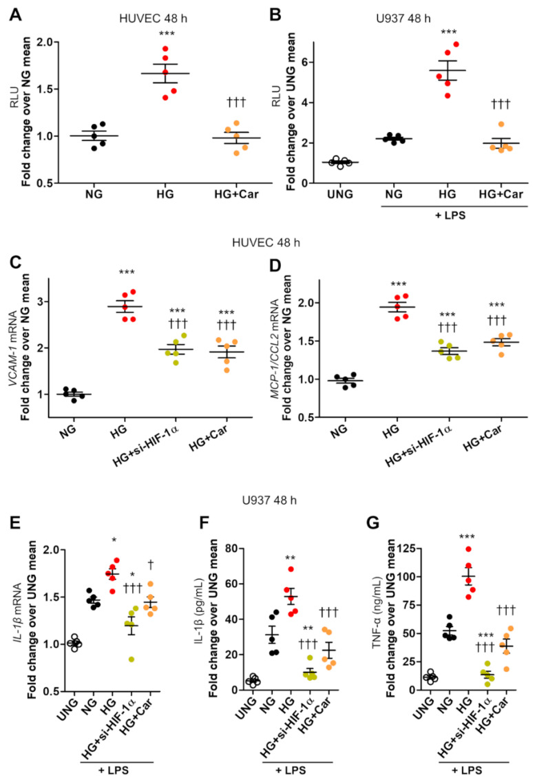
Figure 2.
HG-induced HIF-1α activity is associated with proinflammatory activation in endothelial cells and LPS-stimulated macrophages. HIF-1 activity, as assessed by dual-luciferase gene reporter assay, in HUVEC (A), and in U937 macrophages stimulated (or not, UNG = untreated normal glucose) with LPS (10 ng/mL) (B), after 48 h incubation with HG (20 mM) vs. NG (5.5 mM), with or without Car (20 mM); n = 5 wells in duplicate per condition. VCAM-1 (C) and MCP-1/CCL2 (D) mRNA levels in HUVEC, and IL-1β mRNA levels in U937 macrophages stimulated (or not, UNG) with LPS (E), exposed to HG vs. NG for 48 h, silenced for HIF-1α (si-HIF-1α), or treated with Car; n = 5 wells in duplicate per condition. IL-1β (F) and TNF-α (G) protein levels in the culture medium of U937 cells stimulated (or not, UNG) with LPS after 48 h incubation with HG vs. NG, silenced for HIF-1α, or treated with Car; n = 5 wells in duplicate per condition. Each dot represents the mean of two individual technical replicate and bars represent mean ± SEM. Post hoc multiple comparison: *** p < 0.001, ** p < 0.01 or * p < 0.05 vs. NG; ††† p < 0.001 or † p < 0.05 vs. HG.

