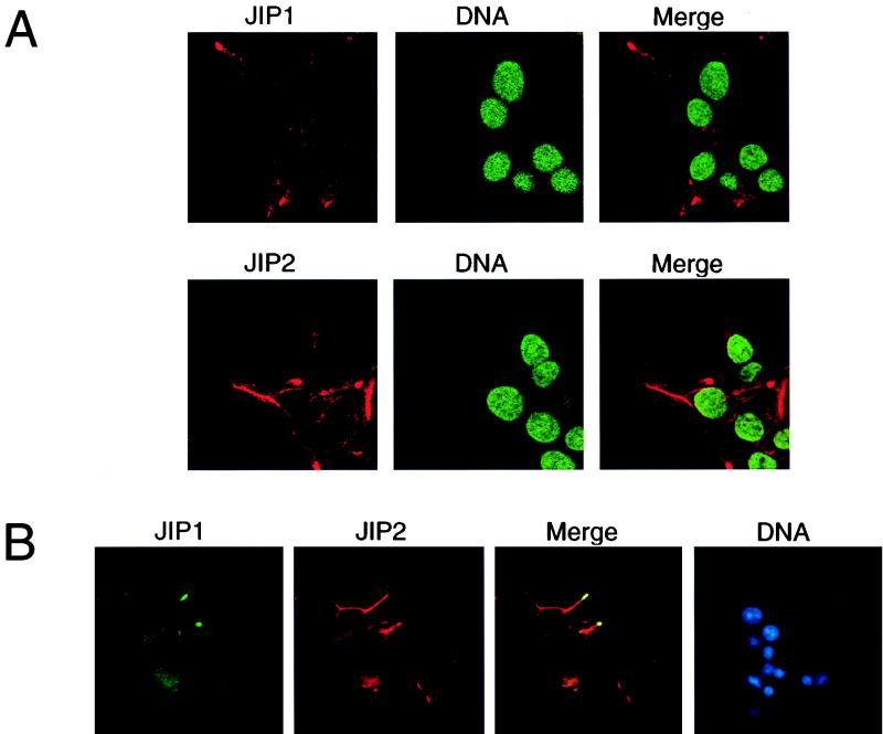
FIG. 10.
Cytoplasmic location of endogenous JIP proteins. (A) The endogenous JIP1 and JIP2 proteins expressed by Rin5F insulinoma cells were detected by confocal immunofluorescence analysis using monoclonal antibodies to JIP1 and JIP2 (red). The nucleus was detected by staining DNA with SYTOX green. (B) Double-label immunofluorescence analysis of JIP1 (green) and JIP2 (red) was performed by conventional microscopy. The nucleus was detected by staining DNA with 4,6-diamidino-2-phenylindole (blue).

