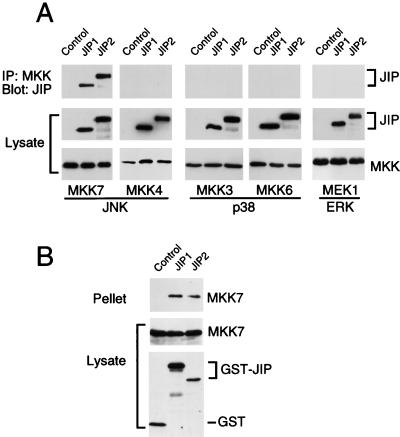
FIG. 3.
Selective binding of JIP1 and JIP2 to the MAPKK MKK7. (A) Epitope-tagged JIP1 and JIP2 were expressed in cells together with epitope-tagged MEK1, MKK3, MKK4, MKK6, or MKK7. Control experiments were performed by transfection of the empty expression vector instead of the JIP expression vector. The expression of JIP and MAPKK proteins was examined by immunoblot analysis of the cell lysates. The MAPKK proteins were immunoprecipitated, and the presence of JIP1 and JIP2 in the immunoprecipitates (IP) was examined by immunoblot analysis using a monoclonal antibody that binds the T7-Tag epitope. (B) The COOH-terminal regions of JIP1 (residues 283 to 660) and JIP2 (residues 499 to 824) were expressed in cells as GST fusion proteins together with epitope-tagged MKK7. Control experiments were performed by transfection with the GST expression vector pEBG. The amounts of the GST proteins and MKK7 in the cell lysates were examined by protein immunoblot analysis. The GST fusion proteins were precipitated from cell lysates with glutathione-agarose, and MKK7 present in the pellet was detected by protein immunoblot analysis using an antibody to the Flag epitope.

