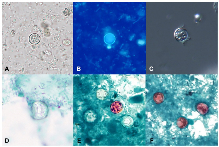
Figure 2.
Oocysts of Cyclospora cayetanensis in stool specimens observed under different staining methods. (A) unstained concentrated wet mount. (B) UV autofluorescence. (C) differential interference contrast (DIC). (D) trichrome stain. (E) Kinyoun’s modified acid-fast. (F) modified safranin. (Figures courtesy of the CDC-DPDx).

