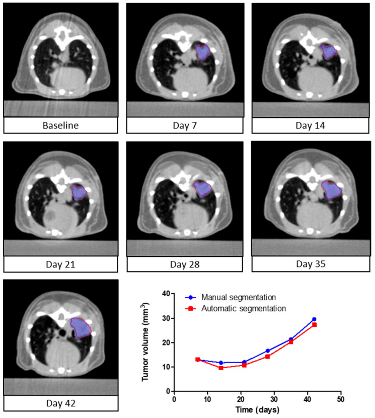
Figure 3.
Longitudinal follow-up of tumor volume. A series of axial slices of µCBCT images showing the manual segmentation (blue) and automatic segmentation (red) of the lung tumor in a single animal versus time. The µCBCT images represent one mouse at seven different time points. The quantified manual (blue) and automatically (red) segmented volumes are presented over time in the line graph.

