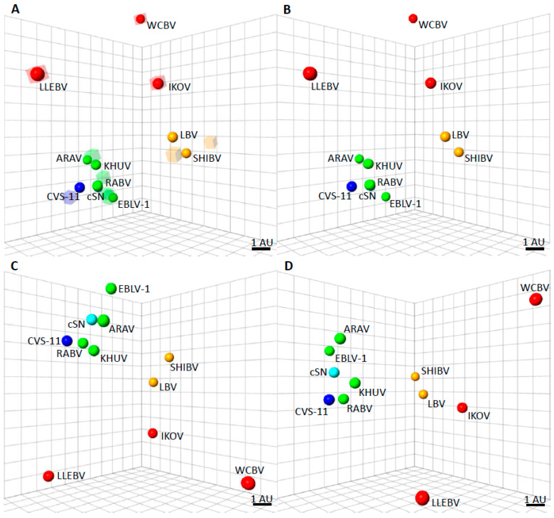
Figure 2.
Antigenic cartography maps to show the antigenic distances of the lyssaviruses. (A) Three-dimensional antigenic map showing the antigenic relationship between lyssaviruses. Viruses (spheres) and, sera (translucent coloured boxes) are positioned such that the distance from each serum to each virus is determined by the neutralisation titre. Multidimensional scaling is used to position both sera and viruses relative to each other, so orientation of the map within the axes is free. The scale bar represents 1 AU (antigenic unit), equivalent to a two-fold dilution in antibody titre. Phylogroup I lyssaviruses are coloured green, CVS-11 coloured dark blue [Challenge virus standard-11 strain of RABV, used routinely in diagnostic assays], cSN [cDNA clone of the SN strain of RABV derived from the RABV strain, SAD B19] coloured light blue, Phylogroup II lyssaviruses coloured orange, and Phylogroup III lyssaviruses coloured red. (B) Antigenic map with sera removed for clarity. (C) Antigenic map, rotated to a different orientation and sera removed for clarity. (D) Antigenic map, rotated to a different orientation and sera removed for clarity.

