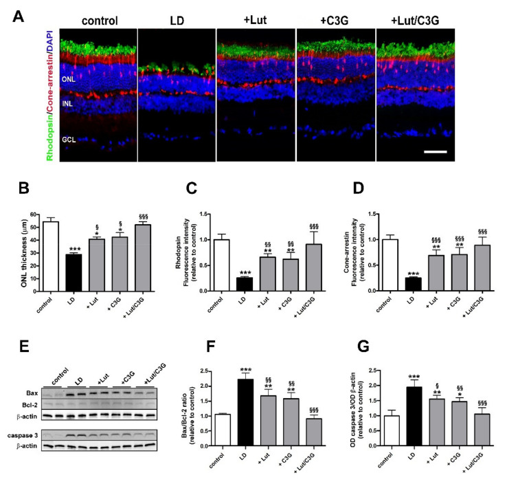
Figure 5.
Individual and combined effects of lutein and C3G on photoreceptor degeneration. (A) Representative images of retinal sections from control and LD rats untreated or pretreated with either lutein, C3G or their combination immunolabeled for rhodopsin (green) and cone-arrestin (red). Retinal nuclear layers are highlighted by the DAPI counterstaining. (B) Quantitative analysis of ONL thickness. (C,D) Quantitative analysis of rhodopsin and cone-arrestin immunofluorescence intensity. GCL, ganglion cell layer; INL, inner nuclear layer; ONL, outer nuclear layer. Scale bar, 50 µm (n = 6 retinas per group). (E) Representative Western blots of Bax, Bcl-2 and active caspase 3 from each experimental group. (F,G) Densitometric analysis of the Bax/Bcl-2 ratio and active caspase 3. The expression of active caspase 3 was relative to the loading control β-actin. Data are expressed as mean ± SD. Differences between groups were tested for statistical significance using one-way ANOVA followed by the Newman-Keuls multiple comparison post-hoc test. * p < 0.05; ** p < 0.01; *** p < 0.001 versus control; § p < 0.05; §§ p < 0.01; §§§ p < 0.001 versus LD.

