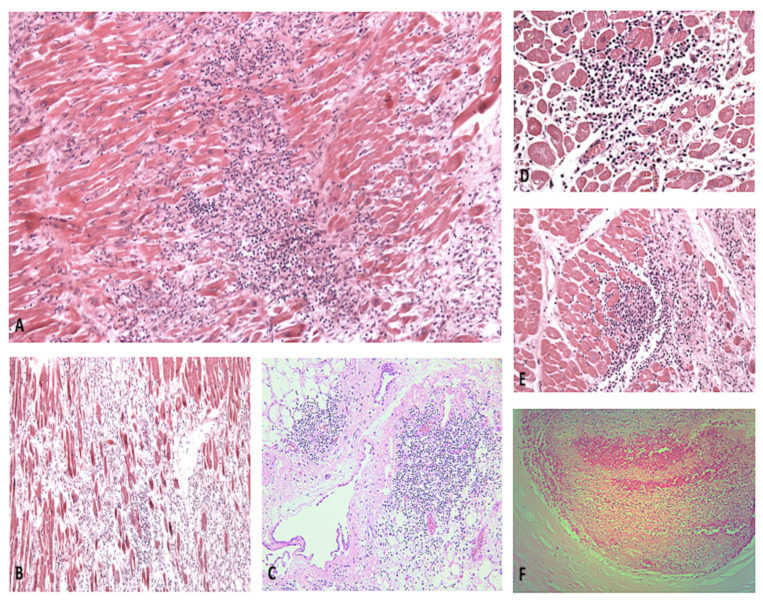
Figure 3.
(A,B) Myocarditis with extensive myocyte necrosis, at different developmental stages (H&E, 60× and 20×, respectively). (C) Massive lymphocytic infiltration of the pericardium and perivascular area (H&E, 40×). Foci of acute myocyte necrosis (D) and regions undergoing tissue repair (E) (H&E, 100× and 80×, respectively). (F) Acute coronary thrombosis (H&E, 200×).

