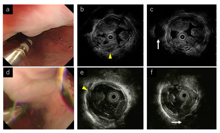
Figure 3.
Endoscopic ultrasound (EUS) images of representative cases. (a) Endoscopic image during the jelly-filling method. (b,c) EUS images of the jelly-filling method. Yellow arrow head shows a perforating vein (Pv). White arrow shows a para-esophageal vein (Para-v). (d) Endoscopic image during the water-filling method. (e,f) EUS images of the water-filling method. Yellow arrow head shows a perforating vein (Pv). White arrow shows a para-esophageal vein (Para-v).

