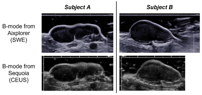
Figure 1.
Example of the B-mode images along the longitudinal plane obtained with the Aixplorer ultrasound imaging system (used for ShearWave Elastography (SWE) imaging) and acquired at matched positions with the Sequoia system (used for dynamic Contrast-Enhanced Ultrasound (CEUS) imaging) for two different subjects. For the two imaging systems the distance between major tick marks is equal to 1 cm.

