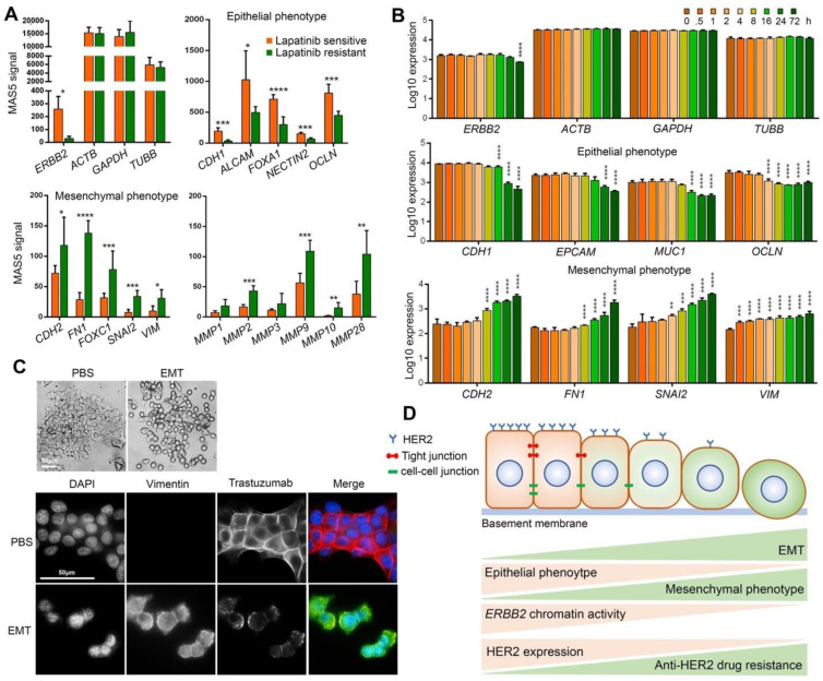
Figure 3.
EMT induces trastuzumab resistance by downregulating HER2 expression. (A) mRNA expression levels of ERBB2 and housekeeping genes, epithelial phenotype marker genes, mesenchymal phenotype marker genes and matrix metalloproteinases in lapatinib sensitive and resistant BT474 cells. (B) mRNA expression levels of ERBB2 and housekeeping genes, epithelial phenotype marker genes and mesenchymal phenotype marker genes in TGF-β-mediated EMT-induced A549 cells. * p < 0.05; ** p < 0.01; *** p < 0.001; **** p < 0.0001. (C) Epithelial morphology of PBS-treated BT474 cells and mesenchymal morphology of EMT-induced BT474 cells (top). Immunofluorescence staining of Vimentin and trastuzumab in EMT-induced BT474 cells treated with 10 μg/mL trastuzumab for 1 h (Bottom). (D) Schematic summary of findings. EMT of HER2-positive breast cancer cells increases trastuzumab resistance by chromatin-based epigenetic downregulation of HER2 expression. Increased EMT and mesenchymal phenotype is correlated with decreased expression of epithelial phenotype (including tight junctions and cell-cell junction proteins) and increased mesenchymal phenotype, decreased chromatin accessibility/activity including reduced enrichment of open/active chromatin marks (H3K4me and H3Kac as examples), increased enrichment of closed/inactive chromatin marks (H3K9me as an example), decreased HER2 expression and eventually increased resistance to anti-HER2 drugs including trastuzumab.

