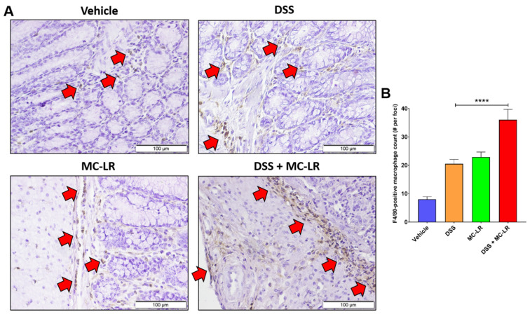
Figure 1.
F4/80-positive macrophages in FFPE colonic sections of DSS-induced colitis model C57BL/6J mice. (A) IHC staining in: (Vehicle) control animals without DSS-induced colitis or MC-LR exposure. (DSS) DSS-induced colitis without MC-LR exposure. (MC-LR) MC-LR exposed animals without DSS-induced colitis. (DSS+MC-LR) DSS-induced colitis with MC-LR exposure. Red arrows denote positive F4/80 staining of macrophages. (B) Quantification of F4/80-positive macrophages by count in 10 random foci per animal (n = 3). Significance by one-way ANOVA (p < 0.0001) and **** = p < 0.0001 by Tukey’s multiple comparisons test.

