Figure 5.
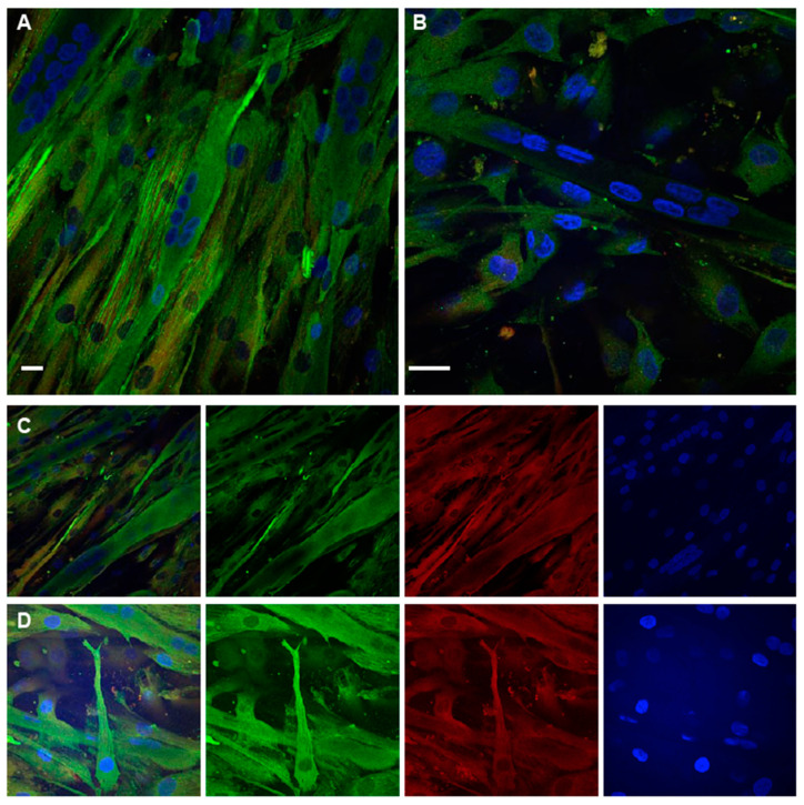
TPM4 and α-actinin staining in differentiating myocytes. (A,B) Confocal immunofluorescence analysis performed at day 1. In the field are present mono-nucleated cells expressing TPM4 (green) organised in filaments and differentiated myotubes with a lower and less organised expression of the protein. (C,D) Panels with separate channels showing the concomitant expression of TPM4 (green) and α-actinin (red) in the cytoplasm. Starting from the left first panel is a merged image of all channels, the second panel shows the captured image using only the 488 nm laser (FITC), the third is the capture using the 561 nm laser (TRITC), the last using the 401 nm laser (DAPI, blue). DAPI is used for nuclei staining. Bars = 10 µm.

