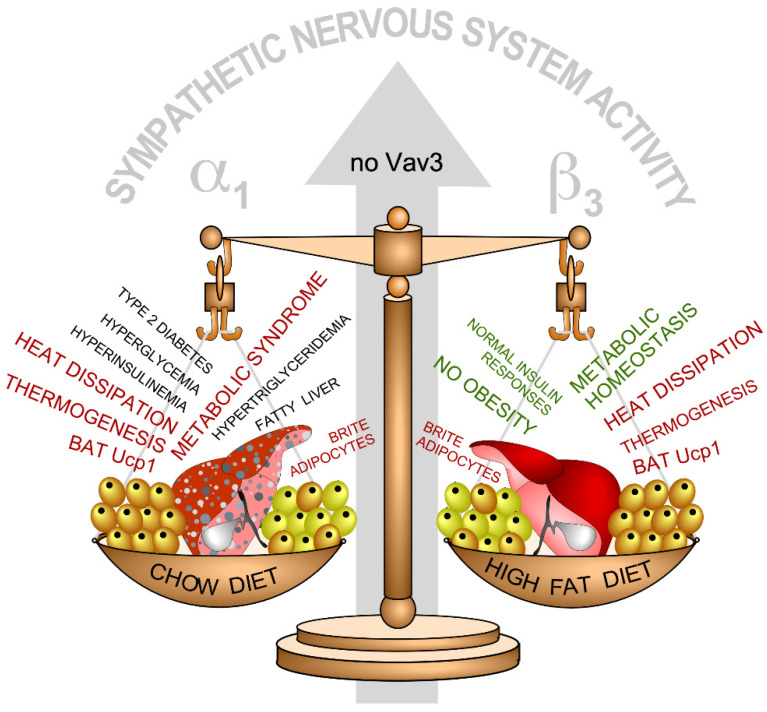
Figure 4.
Summary of the physiological and metabolic dysfunctions found in Vav3–/– mice under chow (left) and high-fat (right) diet conditions in BAT (cells colored in light brown), WAT (cells colored in yellow), liver (red), and plasma. Defects are shown in red and black. Normal responses are shown in green. The main adrenergic receptors involved in the responses found in chow (α1) and high-fat diet (β3) conditions are indicated. Further information is in the main text and reference [63].

