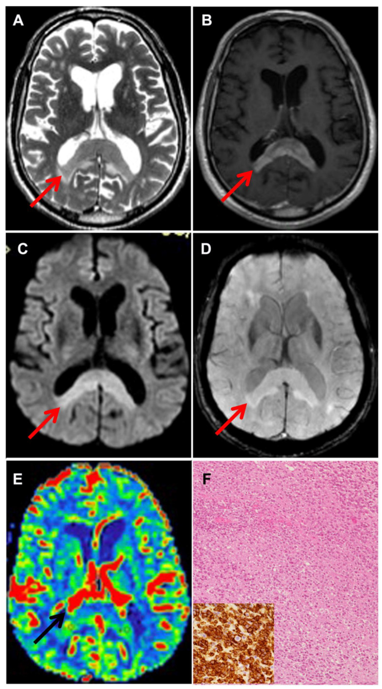
Figure 1.
Neuroradiological and histological features of PCNSL. (A) Axial T2-weighted and (B) axial Gd-enhanced T1-weighted MR imaging showing a T2 hypointense mass involving splenium of the corpus callosum, without necrosis, and with vivid contrast enhancement. DWI (C) showing restricted diffusion and SWI (D) showing absence of intratumoral hemorrhage. Arrows point at the tumor (A–E). CBV map (E) shows moderately increased intratumoral rCBV. The neuropathological examination reveals a high-grade diffuse B cell non-Hodgkin lymphoma (F) showing strong CD20 positivity (F, inset).

