Figure 1.
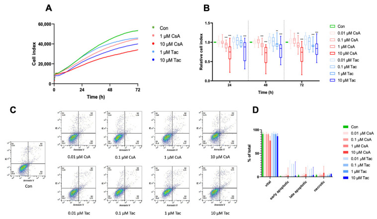
High-dose cyclosporine A (CsA) and tacrolimus (Tac) impaired ECFC proliferation. (A) Overlay of growth curves of ECFCs treated with CsA or Tac (1 µM or 10 µM). (B) ECFC proliferation was not affected after treatment with 0.01 or 0.1 µM CsA or Tac, but it was significantly decreased after treatment with 1 µM or 10 µM CsA or Tac after 24 h, 48 h, and 72 h. n = 13–27; control group set as 1. (C) Representative measurement of apoptosis and necrosis in ECFCs after 48 h treatment with CsA or Tac at 0.01 µM, 0.1 µM, 1 µM, or 10 µM. Viable cells are located in the lower left field (Annexin V neg./PI neg). (D) There was no significant increase of apoptotic or necrotic cells. n = 5. Con—control; CsA—cyclosporine A; Tac—tacrolimus. ** p < 0.01, *** p < 0.001.

