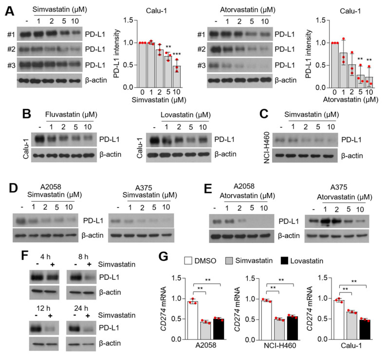
Figure 2.
PD-L1 is decreased in statins-treated lung cancer and melanoma cells. (A) PD-L1 expression in simvastatin and atorvastatin-treated Calu-1 lung cancer cells. Calu-1 cells were incubated with simvastatin and atorvastatin at the indicated concentration for 12 h. DMSO was used for vehicle control (-). PD-L1 protein levels were measured by using western blotting and PD-L1 protein intensity was both quantified and represented by Image J and Graphpad Prism. Each PD-L1 protein level in the statins-treated sample was compared to the vehicle sample. The values represent the mean ± SD for the three independent experiments (#1, #2, and #3) performed. ** p < 0.01 and *** p < 0.001 by one-way ANOVA, followed by an appropriate post hoc test for the comparison between two experimental groups. (B) PD-L1 expression in fluvastatin and lovastatin-treated Calu-1 lung cancer cells. Calu-1 cells were incubated with fluvastatin and lovastatin at the indicated concentration for 12 h. (C) PD-L1 expression in simvastatin-treated NCI-H460 lung cancer cells. Different concentrations of simvastatin were treated for 12 h, as indicated. (D,E) PD-L1 expression in simvastatin (D) or atorvastatin (E)-treated A375 and A2058 melanoma cells. Different concentration of statins were treated for 12 h. (F) PD-L1 expression in simvastatin-treated A2058 melanoma cells in a time-dependent manner, as indicated. (G) CD274 mRNA-encoding PD-L1 protein expression in simvastatin (5 µM for 8 h) and lovastatin (5 µM for 8 h)-treated A2058, NCI-H460, and Calu-1 cells. CD274 mRNA was measured by using qRT-PCR. Values represent mean ± SD. Experiments were performed in triplicates. ** p < 0.01 by one-way ANOVA, followed by an appropriate post hoc test for the comparison between two experimental groups.

