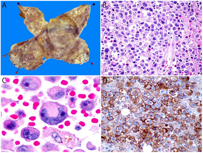
Figure 16.
Breast implant-associated ALCL. (A) Opened breast implant capsule pinned to a flat surface (after overnight fixation). Note the multiple areas with a fibrinous/necrotic appearance on the luminal surface that correspond to collections of lymphoma cells. (B) The cell block obtained from the effusion contains numerous anaplastic large lymphoma cells. (C) At high magnification, the large lymphoma cells show a pleomorphic and hypechromatic nucleus with abundant eosinophilic cytoplasm. A “hallmark” cell is also seen (bottom). (D) The CD30 immunostain shows membranous and paranuclear dot (Golgi) labelling in virtually all these cells.

