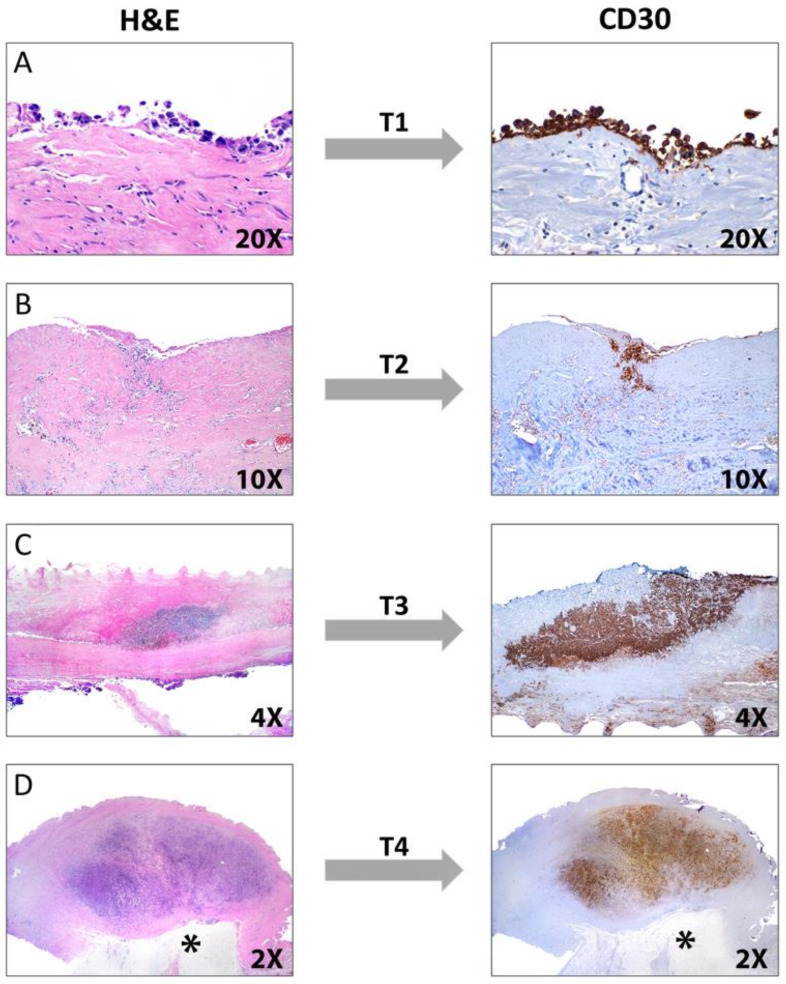
Figure 18.
Pathologic tumor stage (T) used in breast implant-associated ALCL. Left panel: hematoxylin and eosin stain, H&E. Right panel: CD30 immunostain. (A) T1, the lymphoma cells are present only in the effusion and/or confined to the lumen of the fibrous capsule. (B) T2, sparse neoplastic cells infiltrate the capsule. (C) T3, solid aggregates or sheets of lymphoma cells within the capsule; (D) T4, a solid nodule of lymphoma cells occupies the entire capsule thickness and approaches the adipose tissue and breast parenchyma (asterisk). Immunohistochemistry for CD30 is more accurate to evaluate the level of infiltration of the capsule.

