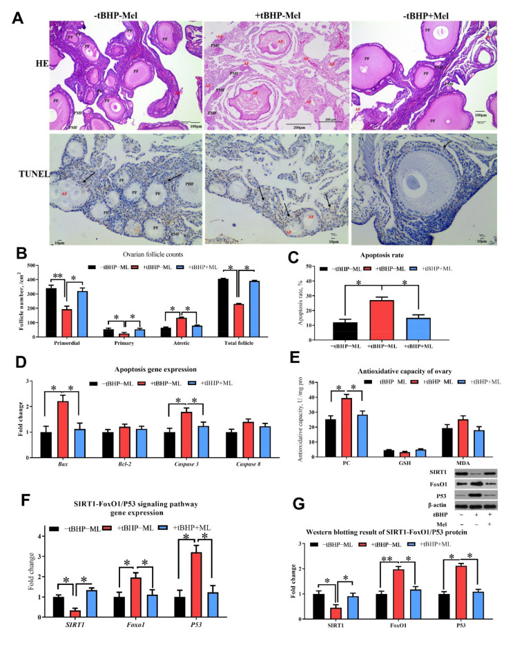
Figure 9.
Melatonin ameliorates oxidative stress (tBHP) induced ovarian dysfunction in vitro (Experiment 2). (A,B) Ovarian histology and TUNEL analysis of layers after three days of treatment and follicle counts at each developmental stage (PMF = primordial follicle; PF = primary follicle; AF = atretic follicle). TUNEL analysis for cell apoptosis in ovary (A,C) showed the TUNEL results with the brown color presents the positive cells. (D) RT-PCR analysis for mRNA expression levels related to ovarian apoptosis gene expression. (E) Antioxidant capacity analysis for ovary with oxidation product. (F,G) the western blotting result of apoptosis associate protein (Bax, Bcl-2, caspase 3 and cleaved-caspase 3). Data are shown as averages and error bars represent SEM± SEM; statistically significant differences are shown with different letters (p < 0.05) with one-way ANOVA with Tukey’s test. −tBHP − Mel = 0 tBHP + 0 melatonin, +tBHP − Mel = 50 μmol/mL tBHP, +tBHP + Mel = 50 μmol/mL tBHP + 100 ng/mL melatonin, Bcl-2 = B-cell lymphoma-2, SIRT1 = silent information regulator 1, FoxO1 = forkhead box O1, PC = protein carbonyl, GSH = glutathione, MDA = malondialdehyde. Statistical significance was evaluated by t-test, * p < 0.05, ** p < 0.01.

